Mastering minimal-sedation endoscopy is not about the scope alone; it’s about deploying a holistic system of patient-centric techniques that transform the procedure.
- Effective patient comfort hinges on micro-decisions, from the physics of insufflation gas (CO2 vs. air) to the psychology of pre-operative sensory environments.
- Strategic choices in bowel preparation for elderly patients and the selective use of disposable scopes for high-risk profiles are as critical as insertion technique.
Recommendation: Adopt an integrated approach that combines superior technology with evidence-based practices in patient preparation, procedural execution, and economic analysis to redefine the standard of care in patient experience.
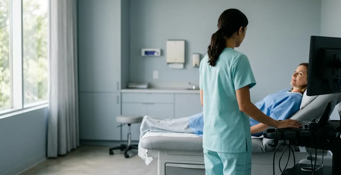
As physicians, we know the look. The tightened jaw, the white-knuckled grip on the gurney—the silent broadcast of anxiety that precedes an endoscopic procedure. For years, the industry’s answer has been primarily technological: smaller, more flexible, ultra-thin scopes. This is a crucial advancement, to be sure. It allows us to navigate the delicate internal architecture of the human body with unprecedented agility, often reducing the need for deep sedation.
However, focusing solely on the hardware misses the bigger picture. We’ve all seen cases where even the most advanced scope can’t overcome poor bowel prep, or where a patient’s anxiety makes a minimal-sedation procedure an ordeal for everyone involved. The platitudes about « patient comfort » are easy to say, but true execution is an art form, a clinical discipline built on a foundation of subtle, interconnected choices.
What if the true key to mastering the « gentle scope » isn’t just the tool in our hands, but the entire system we build around it? This is the core of our discussion. We propose that achieving exceptional patient comfort with minimal sedation is a holistic practice. It’s a synthesis of physiology, psychology, technique, and even economics. It’s about understanding why CO2 is metabolically superior to air, how a split-dose prep can transform compliance in the elderly, and when the hidden costs of reprocessing a reusable scope make a disposable one the wiser choice.
This guide moves beyond the spec sheet of the latest endoscope. We will explore the evidence-based strategies and nuanced clinical decisions that, when combined, create an environment of safety, trust, and genuine comfort for our patients. It’s about transforming the patient journey from a dreaded necessity into a well-managed, minimally disruptive clinical experience.
To navigate this holistic approach, we will delve into the critical components that contribute to a truly patient-centric practice. The following sections break down the key decisions and techniques, from procedural specifics to practice management, that empower us to provide the highest standard of care.
Summary: The Art of the Gentle Scope: Mastering Interior Exams with Ultra-Thin Technology and Minimal Sedation
- Why Using CO2 Instead of Air Insufflation Reduces Recovery Pain by 60%?
- How to Improve Bowel Prep Compliance in Elderly Patients?
- Capsule Endoscopy or Traditional Colonoscopy: Which Is Better for Diagnosing Crohn’s?
- The Insertion Technique Mistake That Increases Perforation Risk in Diverticulosis
- When to Schedule Surveillance Scopes: Adhering to New Polyp Guidelines
- When to Use Disposable Scopes: 3 High-Risk Patient Profiles
- How to Reduce Pre-Op Anxiety Using Sensory Room Modifications?
- Why the Hidden Costs of Cleaning Reusable Scopes Add $50 Per Procedure?
Why Using CO2 Instead of Air Insufflation Reduces Recovery Pain by 60%?
The choice between CO2 and ambient air for insufflation may seem like a minor detail, but it has profound implications for post-procedural patient comfort. While both can achieve luminal distention, their physiological behavior is dramatically different. Air, composed primarily of nitrogen, is poorly absorbed by the colonic mucosa. This trapped gas leads to lingering abdominal distention, cramping, and bloating that can last for hours, significantly impacting the patient’s recovery experience and perception of the procedure.
In contrast, carbon dioxide (CO2) is absorbed 150 times faster than nitrogen. This rapid absorption is not passive; it’s facilitated by the enzyme carbonic anhydrase in the gut wall, which converts CO2 into bicarbonate. The gas is then transported in the blood and efficiently expelled through respiration. This physiological process means that by the time the patient is in the recovery room, most of the insufflated CO2 has already been eliminated from their system. The result is a dramatic reduction in post-procedure pain, a faster return to normal activity, and higher patient satisfaction. In some settings, this can even translate to improved room turnover times, as patients are ready for discharge sooner.
While the benefits are clear, it is crucial to screen for the rare contraindications, such as patients with severe, uncompensated COPD and baseline hypercapnia, who may have a reduced capacity to clear the additional CO2 load. For the vast majority of patients, however, CO2 insufflation represents a simple, evidence-based switch that directly enhances the « gentle scope » experience.
Action Plan: CO2 Insufflation Implementation
- Verify CO2 insufflator compatibility with existing endoscopy equipment.
- Ensure staff understands the carbonic anhydrase enzyme conversion mechanism to explain the rapid CO2 absorption.
- Monitor for a reduction in room turnover time, as 10-15 minutes saved per procedure is a typical outcome.
- Screen for contraindications, specifically severe uncompensated COPD with baseline hypercapnia.
- Document patient pain scores at 1, 3, and 6 hours post-procedure to quantify the improvement.
Ultimately, adopting CO2 is a foundational step in building a practice centered on minimizing patient discomfort from the inside out.
How to Improve Bowel Prep Compliance in Elderly Patients?
Inadequate bowel preparation is a primary cause of cancelled procedures, prolonged procedure times, and missed pathology. This challenge is often magnified in elderly patients, who may struggle with the large volumes, electrolyte shifts, and physical demands of traditional prep regimens. However, the assumption that poor prep is inevitable in this population is a misconception. The key is shifting from a one-size-fits-all approach to a tailored, supportive strategy centered on low-volume, split-dose protocols.
Modern low-volume preparations have revolutionized our ability to achieve excellent cleansing with greater patient tolerance. In fact, dedicated studies show that low-volume preparations achieve a 95.9% adequate preparation rate in the elderly, debunking the myth of guaranteed poor compliance. The split-dose method—consuming half the prep the evening before and the second half the morning of the procedure—is critical. It improves efficacy by cleansing residual stool just before the exam and is generally better tolerated than consuming a large volume at once.
Case Study: Success with Split-Dose Low-Volume Preparation
A significant multicenter study highlighted the effectiveness of this approach. When comparing a split-dose oral sulfate solution (OSS) to a traditional 4L PEG regimen in patients aged 65-75, the results were striking. While both achieved comparable bowel cleansing, the OSS group reported dramatically higher satisfaction and a much greater willingness to repeat the procedure (92.8% vs. 67.7%). This demonstrates that we can achieve our clinical goal of a clean colon without sacrificing the patient’s comfort and cooperation.
Improving compliance also requires clear, large-print instructions, patient education on the importance of the prep, and involving caregivers when necessary. For the patient, a better prep experience is a cornerstone of a gentle procedure.
By prioritizing patient tolerance, we not only improve the quality of our examinations but also reinforce the trust and confidence our elderly patients place in our care.
Capsule Endoscopy or Traditional Colonoscopy: Which Is Better for Diagnosing Crohn’s?
The question of whether capsule endoscopy or traditional colonoscopy is « better » for diagnosing Crohn’s disease is a false dichotomy. A more clinically astute question is: « Which tool is right for which part of the diagnostic journey? » The answer lies in understanding their complementary strengths and limitations. They are not rivals, but partners in achieving a comprehensive diagnosis.
As leading research summarizes, the dynamic is clear. In their « Capsule Endoscopy in Inflammatory Bowel Disease Review, » researchers state:
Colonoscopy is the first-line tool for its therapeutic capability (biopsies), but capsule endoscopy is the superior tool for assessing the vast small bowel, where up to 70% of Crohn’s inflammation can exclusively occur
– Research findings, Capsule Endoscopy in Inflammatory Bowel Disease Review
This single statement perfectly frames the issue. Colonoscopy is indispensable. Its ability to obtain tissue samples for histological confirmation is the gold standard for definitive diagnosis. It also allows for therapeutic interventions like stricture dilation. However, its reach is limited to the colon and the terminal ileum. This is where capsule endoscopy finds its crucial role. Given that a significant portion of Crohn’s disease manifests in the small bowel, the capsule provides a non-invasive method to visualize meters of mucosa that are otherwise inaccessible to a standard colonoscope. This makes it an unparalleled tool for assessing the extent of the disease and for diagnosing patients whose symptoms are suggestive of Crohn’s but who have a negative colonoscopy.
This table from a comparative analysis of diagnostic modalities for Crohn’s disease clarifies the specific trade-offs:
| Feature | Capsule Endoscopy | Traditional Colonoscopy |
|---|---|---|
| Small bowel visualization | Complete (70% of Crohn’s occurs here) | Limited to terminal ileum |
| Biopsy capability | No | Yes – essential for diagnosis |
| Sensitivity for small bowel | 71% detection rate | 65% detection rate |
| Patient comfort | Non-invasive, no sedation | Requires sedation |
| Stricture assessment | Requires patency capsule first | Direct visualization and dilation possible |
The art, therefore, is not in choosing one over the other, but in knowing precisely when to deploy each to build a complete diagnostic picture.
The Insertion Technique Mistake That Increases Perforation Risk in Diverticulosis
Navigating a colon riddled with diverticulosis presents a unique challenge. The accordion-like folds and fixed, angulated segments can easily trap the tip of the endoscope. The most common and dangerous mistake in this scenario is an over-reliance on axial force, or « pushing. » When an endoscopist encounters resistance and simply pushes harder, they are not advancing the scope through the lumen but are instead stretching the bowel wall over the fixed tip. In a segment weakened by diverticular disease, this dramatically increases the risk of barotrauma and perforation.
The solution lies in shifting from a mindset of pushing to one of steering and repositioning. Instead of brute force, expert technique involves using torque (right/left rotation of the scope shaft) to guide the tip around corners and resolve loops. This requires a delicate touch and a constant reading of the visual and tactile feedback from the scope.
Another powerful technique, particularly in a difficult sigmoid colon, is the water immersion method. By instilling water instead of air, the colon becomes heavier and less prone to looping. The water opens up the lumen ahead of the scope, providing a clearer path and reducing the need for forceful advancement. This gentle, hydrodynamic approach aligns perfectly with the philosophy of a minimal-sedation, patient-centric procedure.
Action Plan: Safe Scope Navigation in Diverticular Disease
- Apply torque steering (right/left rotation) instead of axial force to resolve loops.
- Use the water immersion technique to weigh down the colon and reduce looping, especially in the sigmoid.
- Recognize that ultra-thin scopes (e.g., 5mm) are more flexible and may be more prone to looping, requiring technique adjustment.
- Consciously adjust technique when switching between a standard (e.g., 10mm) and an ultra-thin endoscope.
- Monitor for the characteristic accordion-like folds on the monitor, which signal a diverticular segment requiring a more cautious approach.
Switching from force to finesse is not just a safety measure; it’s the hallmark of an expert endoscopist dedicated to minimizing risk and discomfort.
When to Schedule Surveillance Scopes: Adhering to New Polyp Guidelines
For decades, the 5-year interval for post-polypectomy surveillance was a clinical dogma. However, robust long-term data has empowered guideline committees to adopt a more nuanced, risk-stratified approach. A key update is the recommendation for longer surveillance intervals for patients with low-risk findings. This is not about reducing care, but about right-sizing it, avoiding the costs and risks of unnecessary procedures for patients who are at minimal risk of interval cancer.
The most significant change is for patients with only 1 or 2 small (less than 10 mm) tubular adenomas. Under the new guidelines, these patients are now candidates for a much longer follow-up period. In fact, new guidelines recommend surveillance intervals of 7-10 years instead of the previous 5-year standard for this low-risk group. This evidence-based extension has a massive impact on reducing the burden of care on both the patient and the healthcare system.
However, this recommendation comes with a critical prerequisite: the index colonoscopy must be of high quality. We can only be confident in extending surveillance intervals if we are certain the initial examination was thorough. This places a significant responsibility on the endoscopist to not only find and remove polyps but also to document the quality of their own procedure. Adherence to key performance indicators is no longer just good practice; it is the foundation upon which safe, extended surveillance intervals are built.
Action Plan: High-Quality Examination Checklist for Extended Intervals
- Achieve and document a cecal intubation rate greater than 95% to ensure a complete examination.
- Ensure adequate bowel preparation is achieved and documented, aiming for a Boston Bowel Prep Score of 6 or higher.
- Meet or exceed established Adenoma Detection Rate (ADR) benchmarks, a key measure of a meticulous exam.
- Document a withdrawal time of more than 6 minutes, as this is strongly correlated with higher ADR.
- Consider implementing AI-assisted polyp detection (CADe) systems, which have been shown to improve ADR and increase confidence in the examination.
By coupling high-quality examinations with intelligent, risk-stratified surveillance, we provide care that is both clinically sound and resource-conscious.
Key takeaways
- True patient comfort is a system: It combines technology (ultra-thin scopes) with technique (CO2, water immersion) and strategy (prep compliance, sensory modulation).
- The right tool for the job: Capsule endoscopy and traditional colonoscopy are complementary, not competitive, particularly in Crohn’s disease diagnosis.
- Quality is paramount: Extending surveillance intervals based on new guidelines is only safe if the index colonoscopy meets high-quality benchmarks like ADR and withdrawal time.
When to Use Disposable Scopes: 3 High-Risk Patient Profiles
The advent of single-use, disposable endoscopes presents a paradigm shift in how we approach infection control and resource management. While not a replacement for reusable scopes in every situation, they offer a powerful solution for specific, high-risk scenarios where the absolute guarantee of sterility is paramount. Moving beyond the general benefit of « no cross-contamination, » a strategic clinician thinks in terms of patient profiles. There are three clear profiles where the use of a disposable scope is not just a convenience, but a critical patient safety measure.
The first profile is the immunocompromised patient. This includes individuals on biologics for Inflammatory Bowel Disease (IBD), transplant recipients, or patients undergoing chemotherapy. For them, even a low-level exposure to pathogens that might be harmless to a healthy individual can trigger a serious infection. The guaranteed sterility of a disposable scope eliminates this risk entirely.
The second profile is the patient with known colonization by a multidrug-resistant organism (MDRO). This includes organisms like Carbapenem-resistant Enterobacteriaceae (CRE), Vancomycin-resistant Enterococci (VRE), or Clostridioides difficile (C. diff). Using a reusable scope on such a patient, even with perfect reprocessing, carries a small but non-zero risk of subsequent transmission. Using a disposable scope contains the threat and protects the entire patient population, as well as the equipment itself from complex decontamination protocols.
The third, and often overlooked, profile is the emergency, after-hours procedure. Scenarios like an urgent GI bleed or an emergent ERCP often occur when the full, experienced sterile processing team is unavailable. In this high-pressure environment, a disposable scope ensures perfect, out-of-the-box sterility without delay, removing a significant variable from a complex clinical situation.
In these cases, the disposable scope is not just an alternative; it is the superior clinical choice, prioritizing patient safety above all else.
How to Reduce Pre-Op Anxiety Using Sensory Room Modifications?
Patient anxiety is a physiological reality that can complicate even the most technically straightforward procedure. It can increase heart rate, elevate blood pressure, and heighten the perception of pain, often necessitating higher levels of sedation. While verbal reassurance is important, a more powerful approach is to proactively engineer a calming environment through sensory modulation. This involves intentionally modifying the auditory, tactile, and visual inputs the patient receives before the procedure even begins.
This strategy moves beyond simply trying to distract the patient and instead aims to actively down-regulate the nervous system. Simple, low-cost interventions can have a dramatic effect. For example, studies show that virtual reality distraction with VR goggles, even in pediatric patients, can be highly successful. Immersive nature scenes can transport a patient from a sterile clinical room to a tranquil beach, effectively interrupting the feedback loop of anxiety.
The most effective approach is to have a « Mobile Sensory Modulation Toolkit » available. This allows staff to tailor interventions to the individual patient’s needs, creating a personalized cocoon of calm. This is not about extravagant spa-like rooms, but about a practical toolkit that can be deployed at the bedside.
Action Plan: Mobile Sensory Modulation Toolkit Components
- Provide noise-cancelling headphones with options for binaural beats or calming music.
- Offer weighted blankets, which provide deep pressure stimulation shown to have a grounding, calming effect.
- Set up portable aromatherapy diffusers with calming scents like lavender or chamomile.
- Implement and train staff on verbal sedation and hypnotic language coaching scripts to guide the patient’s focus.
- Deploy VR headsets pre-loaded with peaceful nature scenes (e.g., beaches, forests).
- Train staff in hypnotic language techniques to guide patients into a state of relaxation.
This patient-centric focus transforms the waiting period from a time of stress into the first step of a smooth, gentle procedure.
Why the Hidden Costs of Cleaning Reusable Scopes Add $50 Per Procedure?
The upfront cost of a reusable endoscope is just the tip of the iceberg. The true cost of ownership becomes clear only when we account for the extensive, resource-intensive process of reprocessing. While the per-procedure cost of a disposable scope seems high at first glance, a detailed breakdown of the « hidden » costs associated with cleaning a reusable scope reveals a much more balanced economic picture. These costs, often buried in departmental budgets, can easily add up to $50 or more for every single cleaning cycle.
These are not abstract figures; they are tangible expenses. They include the consumable materials like enzymatic detergents, specialized brushes, and the personal protective equipment (PPE) for staff. They include the significant utility costs of water and electricity required for automated endoscope reprocessors (AERs). Furthermore, there’s the amortization of the AER itself and the opportunity cost of having a scope out of commission for the 90-plus minutes it takes to complete a full reprocessing cycle. Each of these components contributes to the total per-procedure cost, as detailed in this endoscopy cost analysis.
| Cost Component | Estimated Cost |
|---|---|
| Enzymatic detergents & cleaning solutions | $8-12 |
| Staff PPE per cleaning cycle | $3-5 |
| Water & electricity consumption | $2-4 |
| Brush & channel cleaner replacement | $5-7 |
| AER amortization | $10-15 |
| Scope downtime opportunity cost (90 min) | $15-20 |
| Risk mitigation insurance | Unquantifiable |
However, the most profound insight comes when we move beyond direct costs and consider risk. A single cross-contamination event can lead to catastrophic financial and reputational damage. From this perspective, the higher unit cost of a disposable scope can be viewed as a form of insurance. As one healthcare economics analysis puts it:
While not a direct per-procedure cost, the catastrophic financial and reputational damage of a single cross-contamination event acts as risk-mitigation where the higher per-unit cost of a disposable scope serves as an insurance policy
– Healthcare Economics Analysis, Endoscopy Cost Analysis
This economic reality reframes the debate, forcing us to consider the total cost of providing safe, high-quality patient care, not just the initial purchase price of our tools.